
Bioconvergence
Bioconvergence is a new interdisciplinary scientific research area that combines biotechnology, chemistry, engineering, and computer science to solve complex challenges in healthcare, sustainability, agriculture, and other fields. It enables innovations such as personalized medicine, regenerative therapies, advanced diagnostics, and bio-inspired materials. Bioconvergence has the potential to transform the way we prevent, predict, and treat diseases, as well as improve the quality of life for millions of people around the world. Researchers at BINA are involved in various aspects related to bioconvergence, including:
- Biochips
- Biosensors
- Human Organs-on-chips
- Neuron-chip and cell-chip interfaces
- Optical and electronic micro-devices
- Microfluidics and Lab on chip
- Electrochemical sensors
Researchers
-
The Biofilm Research Laboratory
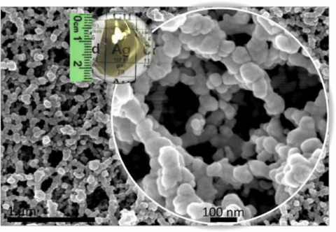
• Bacterial biofilms
• Nanoparticles with anti-biofilm properties
• Bacterial virulence
• Bio-ethanol production -
Prof. Amos Danielli
972-3-738-4653
Optical Imaging and Biosensing Laboratory
• Rapid and highly sensitive detection of biomarkers, such as proteins and specific DNA sequences
• Detection of protein-protein interactions
• Magnetic manipulation of nanoparticles, design of magnetic poles, magnetic force optimization -
Prof. Doron Gerber
972-3-738-4508 -
Prof. Nitzan Gonen
972-3-5317246
Sex Determination
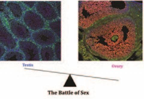
• 3D Genome organisation
• CRISPR genome editing
• Stem Cell Biology
• Developing in vitro systems to model the gonads -
Prof. Gil Goobes
972-3-5317390 -
Prof. Ayal Hendel
972-3-531-7316
Precise and efficient CRISPR genome editing as a curative therapy for genetic disorders
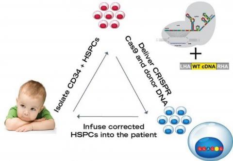
• Biotechnology
• Genetic therapy
• Genetic engineering
• Developing CRISPR technology as a method of gene therapy for genetic diseases -
Prof. Beena Kalisky
972-3-738-4339
Sensitive magnetic imaging
• Superconductivity
• Nano-magnetism
• Bio-magnetism
• Scanning SQUID microscopy
• Complex oxid interfaces
• Nano-electronics -
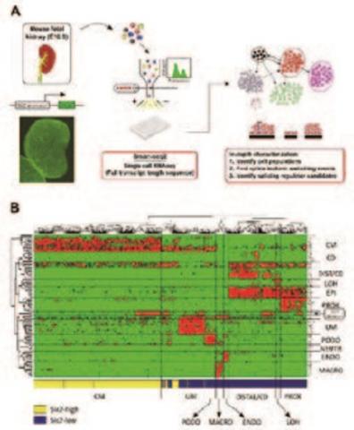
Prof. Tomer Kalisky
972-3-738-4656
Single-cell genomics of kidney development, regeneration, and cancer
• Biochips & Sensors
• Disease Treatment
• Drug Delivery
• Genomics, Proteomics & Glycomics
• Imaging -
Biological network systems
Our research lab is interdisciplinary, dedicated to unraveling the complexities of complex biological network systems, with a particular focus on gene regulatory networks, aging, and human microbiome. We develop innovative computational methodologies drawn from a variety of fields, including artificial intelligence, computational biology, ecology, nonlinear dynamics, statistical mechanics, and especially network science. We rigorously validate our theories and approaches through carefully designed biological experiments conducted in our lab. Our ultimate goal is to enhance our understanding of tissue biology and physiology, leading to improved diagnostics and therapeutics that can significantly advance patient care. Central to our research is the exploration of the intricate relationships between the structure and dynamics of complex networks, which are fundamental to tissue function and stability.
-
Prof. Dan Thomas Major
972-3-531-7392 -
Prof. Yossi Mandel
972-3-738-4234
Ophthalmic Science and Engineering Lab
• Electro-cellular interfaces, optical and electronic micro-devices development
• Applied science for improving diagnosis, treatment and prevention of various ophthalmicdiseases.
• Artificial introduction of the visual information and its processing by the retina and the visual cortex.
• Electro-cellular interface with the autonomic system and application of high electrical field for solid tumor ablation (IRE - Irreversible Electroporation). -
Prof. Shulamit Michaeli
972-3-531-8068
Mechanism and machinery of nucleolar gene silencing
• The use of nanoparticles for cytoplasmic and nuclear gene silencing
• Trans-splicing in trypanosomatids
• Protein translocation in trypanosomatids
• RNA modifications mediated by guide RNAs
• RNAi silencing in plants
• The use of nano particles as RNAi carriers into the nucleous -
Optical and Acoustical Neuroimaging Lab
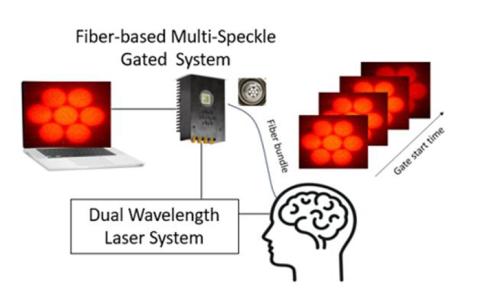
Our neuroengineering research focuses on developing advanced acoustical and optical neuroimaging methods to understand the brain's neural circuitry and fundamental mechanisms. These methods have significant potential in brain-computer interfaces and clinical studies. We combine cutting-edge Electrical Engineering and Neuroimaging techniques, including single photon sensing, superconductive sensing, machine learning, FPGA design, nanoelectronics, biomedical sensing, and neuroimaging.
-
Prof. Rachela Popovtzer
972-3-531-7509
Nanotheranostics for Personalized Medicine
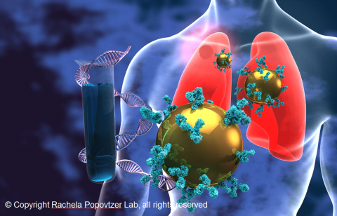
• Molecular CT imaging of cancer using targeted gold nanoparticles.
• Theranostic approaches for Alzheimer’s and Parkinson’s Diseases.
• Metabolic based imaging and therapy.
• Optical/chamical imaging of enzymatic activity.
• Nanoparticle-based strategies for targeted drug delivery.
• In-vivo cell tracking techniques. -
Prof. Sharon Ruthstein
972-3-7384307 -
Prof. Adi Salomon
972-3-738-4235
Light-matter interaction at the nanoscale
The overall goal of my laboratory is to develop, fabricate and to use plasmonic systems as a ‘photonic environment’, or even as a ‘photonic catalyst’. In general, we aim at opening new routes for photochemical processes/reactions on surfaces by controlling the electromagnetic-field properties at the metal surface, that is, to do, 'chemistry with plasmons'.
-
Theoretical and Computational Research on Polymers in Materials and Biophysics
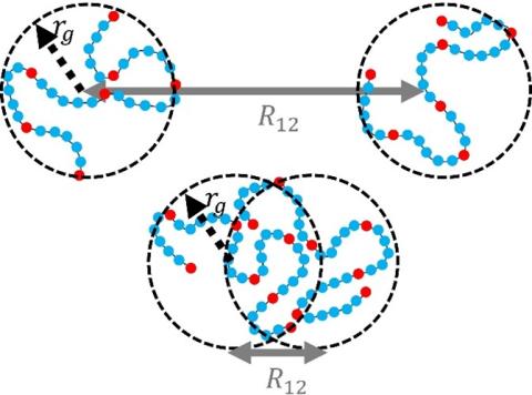
Our research focuses on understanding the structure and dynamics of polymers through theoretical models and computational simulations. In biophysics, we study how intrinsically disordered proteins interact and examine the formation and properties of biopolymer condensates created through liquid-liquid phase separation. In materials science, we investigate how small molecules move through polymer membranes and analyze the elasticity of interpenetrating polymer networks. Throughout our research, we seek to understand the molecular mechanisms behind these phenomena and validate our findings using molecular dynamics simulations. We conduct simulations of polymer solutions, polymer assemblies, and polymer networks, and test the effects of external stimuli on these systems. Our long-term goal is to develop quantitative theories that can predict the properties of polymeric materials and that will eventually allow the rational design of new materials.
-
Prof. Amos Sharoni
972-3-738-4516
Phase transitions on the nano-scale
- Spintronics
- New Temperature Coefficient of Resistance (TCR) materials
- Organic/SC hybrid
-

Prof. Orit Shefi
972-3-531-7079
Neuroengineering and Regeneration
• Neurobiological systems development: image processing and network analysis
• Tissue Engineering: Developing skin grafts that enable reinnervation and regeneration
• Developing devices for reagents delivery into live tissue at a microscopic resolution
• Neuroprosthetic devices: Neuron-Chip interface -
Prof. Yaakov Tischler
972-3-738-4514
In the Device Spectroscopy Laboratory, we use optical spectroscopy to study nanoscale materials such as molecular organics and more generally nanostructured semiconductors, and then devices composed of these materials
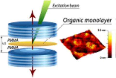
• Coherent coupling in light-matter coupled systems: Organic Lasers, J-aggregates, and Polaritons.
• Ultra-high resolution scanning microcopy and spectroscopy.
• Applications of ultra-fast non-linear spectroscopy for energy sustainability.
• Novel approaches to organic crystal growth and OLED deposition -
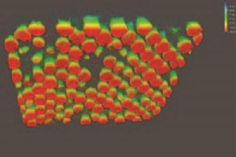
Dr. Moran Yadid
0543561226
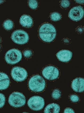
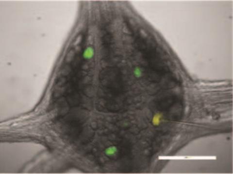
Bioengineering and Regenerative Medicine
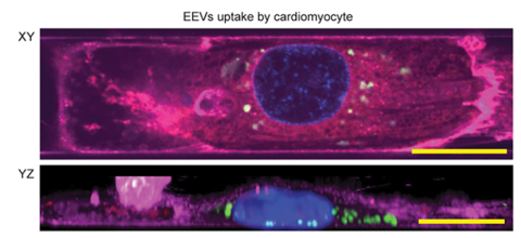
A single cardiomyocyte, micropatterned on soft silicon surface. The images demostrates two views of uptake of fluorescently labeled extracellular vesicles in the cardiomyocyte.
Image was taken by a spinning disk Olympus microscope.
-
Dr. Einat Zalckvar
03-7384013