
Nano CT Service
Non-destructive Tomography
An advanced 3D X-ray nano-tomography platform for non-destructive, multi-scale imaging from millimeter to sub-micron resolution, offering high mechanical and thermal stability for precise quantitative characterization of structural and compositional features in a broad range of materials
BINA's Characterization Unit welcomes both industry professionals and researchers – providing state-of-the-art equipment, expert support, and customized solutions. We’re here for you!
Contacts: Dr. Alexander Domantovsky, alexander.domantovsky@biu.ac.il
Dr. Gili Cohen Taguri, Gili.cohen-taguri@biu.ac.il
Model: SkyScan 2214
Manufacture: Bruker
Specifications:
- Resolution Down to the sub-micron range
- X-Ray Source Adjustable voltage/power for diverse materials
- X-Ray Energy 20–160 kV
- Maximum Power Up to 16 W
- Spot Size 500 nm (LaB6 filament) or 800 nm (W filament)
- Detectors 4-detector array: Flat-panel, CMOS1, CMOS2, and CMOS3
- Max Sample Size Ø 300 mm (140 mm scan area); 400 mm length; 20 kg weight
Applications:
- Materials Science & Engineering: Microstructural characterization of metals, ceramics, polymers, and composite systems.
- Additive Manufacturing: Detection and quantification of sub-micron defects, porosity, and internal cracks in 3D-printed components.
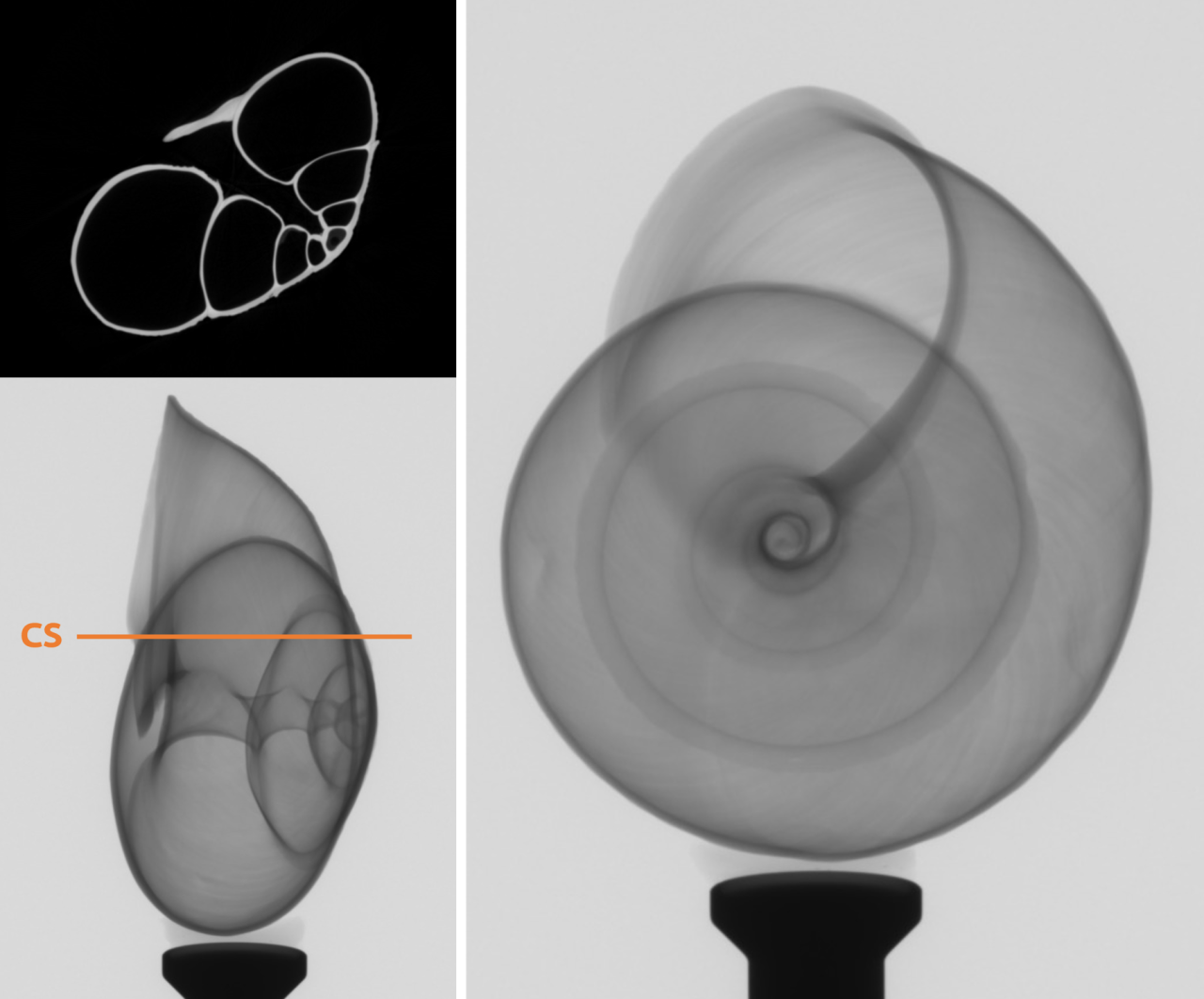
- Life Sciences: 3D imaging of mineralized tissues, soft tissues, plants, and insects.
- Geoscience: Structural analysis of soil, rock, and porous geological materials.
Capabilities:
- Multi-scale imaging: Seamlessly transition from millimeter-scale samples down to sub-micron resolution (500 nm) within a single workflow.
- High mechanical stability: Anti-vibration granite base with pneumatic leveling ensures rotation positioning accuracy below 50nm.
- Material Versatility: Adjustable X-ray source (20-160 kV) allows for the imaging of a wide range of materials, from soft biological tissues to dense metals
- Non-Destructive Testing: enables the study of internal morphology, porosity, cracks, and density variations without the need for physical sectioning
- Quantitative 3D analysis of porosity, defects, phase distribution, and internal interfaces
- Reconstruction and analysis using dedicated tomography software tools