


Spitting images
Until recently, BINA’s Charged Particle Microscopy facility has provided high-resolution atomic imaging and analysis of material sciences samples. Now, due to the expertise of Dr. Ayelet Atkins, BINA has added the capacity of sample preparation and analysis of biological specimens. These new methods serve the industrial and scientific community for advancing bio-medicine, including cancer research, as well as semiconductor research and other nanotechnology applications.
“The TEM (Transmission Electron Microscopy) is a sophisticated microscope that analyzes specimens less than 100 nm thick”, said Dr. Ayelet Atkins, the head scientist in charge of operating the device. The TEM contains an electron gun that sputters electrons towards the sample’s surface, forming a high-voltage beam. As the beam is transmitted through the specimen, the electrons interact with the sample and a 2D high-resolution black and white image is created. The image is suspended on a grid, or a “mesh”, magnified and focused onto an imaging device, such as a fluorescent screen, a sensor or a camera.
Atkins points out that capturing particle images at a very high resolution is achieved due to the small wavelength of electrons in matter (also termed De Broglie waves). “We can view details as small as a single column of atoms, thousands of times smaller than in a light microscope. We can analyse chemical and biological samples, such as viruses and cancer cells, and look at the material’s internal composition, crystallization, stress and magnetic attributes”, she said.
The CPM facility provides services to BIU scientists, as well as for other academic institutions, private companies and hospitals in Israel. A current project is aimed at the discovery of kidney disease. “When viewing deposits on kidney tissue we can measure tissue thickness and identify different types of kidney disease in patients”, Atkins explained.
Dr. Atkins was born in Milford, MA, U.S.A, and moved to Israel 25 years ago. She has wor ked at the CPM facility since January 2017, having gained specialized expertise from her Ph.D. and her postdoc. During her Ph.D. at the Hebrew University and at the Weizmann Institute she researched the material properties of acellular fish bone. “Acellular bone, which exists in most fish species, does not contain osteocytes. When we applied load sensors to this fish bone we found that it has a mechanism that compensates for the lack of osteocytes, and exhibits adaptive, plastic response to loading conditions. The idea was to apply this mechanism for treating patients with skeletal diseases”, she said.
During her postdoc at the University of Toronto Atkins researched the effect of micro-damage accumulation in metastatically-involved vertebrae. The objective was to implement preventive measures in bone cancer patients, by improving fracture risk prediction and bone strength assessment.
“We recently analyzed samples provided by Prof. Ronit Sarid, head of a lab at BINA, using TEM to characterize the function of the herpesvirus (Fig. 1). This is part of a study which Sarid is conducting on Kaposi’s sarcoma-associated herpesvirus (KSHV), a virus known to trigger cancer. The research
aims to shed light on the func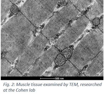 tion and regulation of selected viral genes and their protein products that are potentially involved in the pathogenesis of KSHV”, Atkins explained.
Another project in which Atkins is involved is led by Prof. Haim Cohen from Bar Ilan University, who is researching the aging mechanism and its effects on muscle morphology (Fig. 2). To gain a better understanding of the molecular pathways of aging, the team is investigating caloric restriction and sirtuin proteins−two major factors that have been shown to extend lifespan and delay age-related diseases such as cancer and diabetes.
Got eagle eye-sight?
An additional technique used in structural biology and chemical particle analysis is Cryogenic TEM. Using this method, samples are cooled to cryogenic temperatures of between -170 to-180°C, which halts the movement of the particles and prevents them from aggregating.
SEM is another instrument which provides the ability to view larger samples, and analyze numerous specimens simultaneously. “Using SEM the image we get conveys an illusion of a 3D image, which can be used for studying topography, morphology, chemistry, crystallography and grain orientations”, Atkins said.
“ESEM (environmental scanning electron microscope) is a variation of SEM, used for in-situ  experiments, such as reactions with the atmosphere and temperature effects. A fascinating research project involving the ESEM is currently conducted by Dr. Yossi Mandel’s team at BINA. They are developing a hybrid retinal implant that restores vision in people suffering from retinal degeneration. The implant contains live neurons that are still functional, integrated within an electrode array, to emulate original natural vision (Fig. 3),” described Atkins.
Last Updated Date : 20/04/2021